Ultrasound Molecular Imaging
What if doctors could detect atherosclerosis in at-risk patients before arterial plaques ever develop? What if cancer could be detected and treated at the first sign of unregulated cell growth?
Ultrasound molecular imaging is a powerful imaging method which utilizes microbubble contrast agents to target and bind disease markers in the blood vessels. While traditional imaging methods detect disease by gross anatomical features, we aim to detect disease on a molecular level. This unprecedented level of imaging sensitivity can not only improve the contrast and clarity of diagnostic images, but significantly improve patient outcomes as physicians make faster, more accurate diagnoses. In our lab, we program ultrasound imaging sequences and image processing techniques to improve the contrast and isolation of microbubble signals. Our studies work to advance the field of ultrasound molecular imaging toward rapid clinical adoption.
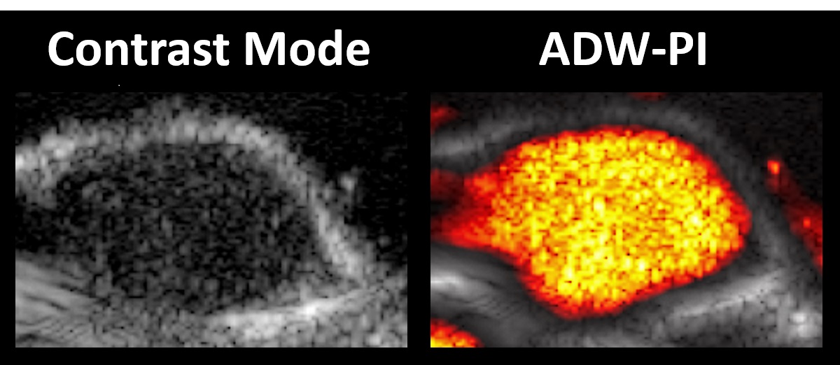
Figure 1. Ultrasound image of a mouse hindlimb tumor with adherent microbubbles on the tumor interior. Microbubble signal was successfully isolated using a decorrelation-based imaging technique developed in Hossack Lab (Herbst, et al. 2017).
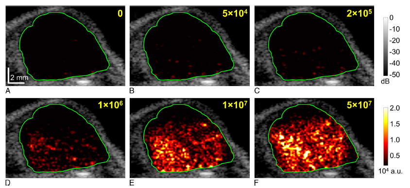
Figure 2. Ultrasound images of a mouse hindlimb tumor administered with different quantities of targeted microbubbles (yellow). High-sensitivity contrast imaging techniques were developed in Hossack Lab and shown to detect microbubble adherence in dosages as low as 5×10^4 microbubbles per injection, representing a 20-fold increase in imaging sensitivity (Wang, et al 2016).